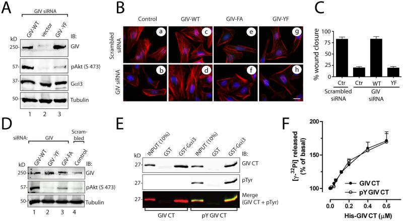
Figure 2. Tyrosine phosphorylation of GIV does not affect its ability to bind or activate Gαi, but is required for phosphorylation of Akt, actin remodeling, and cell migration.
A. HeLa cells stably expressing control vector or siRNA resistant GIV-WT-FLAG or GIV-YF-FLAG plasmids were transfected with GIV siRNA. Lysates were immunoblotted for GIV, phospho-Akt (pAkt), Gαi3, and tubulin. Phosphorylation of Akt (pAkt) was significantly reduced in GIV-depleted control cells (lane 2) and GIV-YF cells (lane 3) compared to GIV-WT cells (lane 1). p < 0.001 for both sets of comparisons; N = 3 experiments. B. HeLa cells stably expressing vector (control), siRNA resistant GIV-WT-FLAG, GIV-FA-FLAG (the GEF-deficient mutant), or GIV-YF-FLAG plasmids were treated with scrambled or GIV siRNA as indicated. Cells were costained with phalliodin-Texas red (F-actin, red) and DAPI (DNA, blue). Depletion of GIV in control cells resulted in loss of stress fibers, which were restored by expression of siRNA resistant GIV-WT-FLAG, but not GIV-FA-FLAG or GIV-YF-FLAG. Bar = 10 μM. C. Untransfected HeLa cells (control; Ctr), HeLa-GIV-WT, HeLa-GIV-FA, and HeLa-GIV-YF cells were transfected with scrambled or GIV siRNA. Results are shown as mean ± S.D. of 8-12 randomly chosen fields from n=3 experiments. p < 0.001 for either sets of comparisons between Ctr and GIV-depleted cells and between GIV-WT-FLAG and GIV-YF-FLAG cells. D. Control HeLa cells and HeLa cells stably expressing siRNA resistant GIV-WT-FLAG, GIV-YF-FLAG, and GIV-FA-FLAG plasmids were transfected with scrambled and GIV siRNA and immunoblotted for GIV, phospho-Akt (pAkt), and tubulin. p -values for both comparisons between GIV-WT-FLAG and GIV-FA-FLAG and between GIV-WT-FLAG and GIVYF-FLAG are < 0.001; N = 3 experiments. (E) Mock-treated His-GIV-CT and in vitro EGFR-phosphorylated His-GIV-CT were incubated with GST-Gαi3 or GST preloaded with GDP immobilized on glutathione beads. Bound proteins were analyzed by two-color immunoblotting (IB) for GIV CT (His) and pTyr. (F) The amount of GTP hydrolyzed in 10 min by His-Gαi3 was determined in the presence of the indicated amounts of sham-treated and in vitro EGFR-phosphorylated His-GIV-CT.

