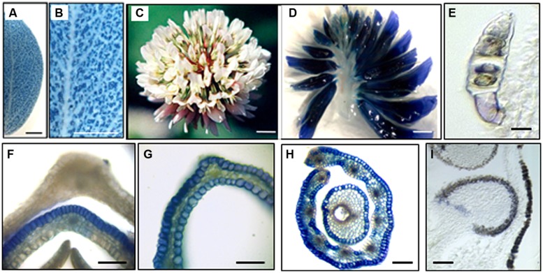
FIGURE 2.
Accumulation of proanthocyanidins in Lotus corniculatus and Trifolium repens organs. Abeynayake et al. (2011,2012). (A,B) L. corniculatus leaf stained with DMACA; (C) mature T. repens inflorescence; (D) longitudinal section through mature T. repens inflorescence stained with DMACA; (E) longitudinal section through a trichome; (F) transverse section through an immature petal in which proanthocyanidins accumulated only on the abaxial side. (G) transverse section through a mature standard petal showing the accumulation of proanthocyanidins on both the abaxial and adaxial sides; (H) transverse section through anther filaments and carpel; (I) longitudinal section through a developing seed in a mature flower. Bars = 200 μm (A,B); 1 mm (C,D); 5 μm (E); 50 μm (F,G), 100 μm (G), and 10 μm (I).

