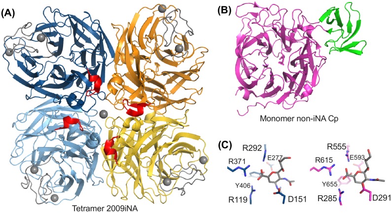
Figure 2.
NA folds of (A) Tetrameric iNA with iNA-specific insertions highlighted in red (insertion before D151) and gray (Ca2+ ion binding site). (B) Bacterial NA of C. perfringens (non-iNA Cp) with the non-catalytical domain in green. (C) Respective active sites with ligands (gray) zanamivir in iNA (blue) and non-iNA Cp (pink). Six conserved active site residues are shown in stick representation. Structures of NAs from further investigated systems are shown in Figure S1.

