
Figure 5.
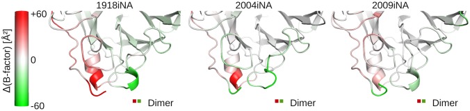
Identification of the 110-helix and the extended 150-loop as assembly sensitive regions based on three ligand-free dimer simulations. The iNA protein backbone is color coded by the differences in B-factors between the two subunits in the dimer. Protein regions colored white indicate areas of similar fluctuation in both subunits. For the 110-helix higher B-factors are observed (red coloring) in the first subunit, where this region is solvent exposed (red in dimer legend). The 150-loop is in the second subunit (green in dimer legend), resulting in negative B-factor differences (green coloring).