Figure 2.
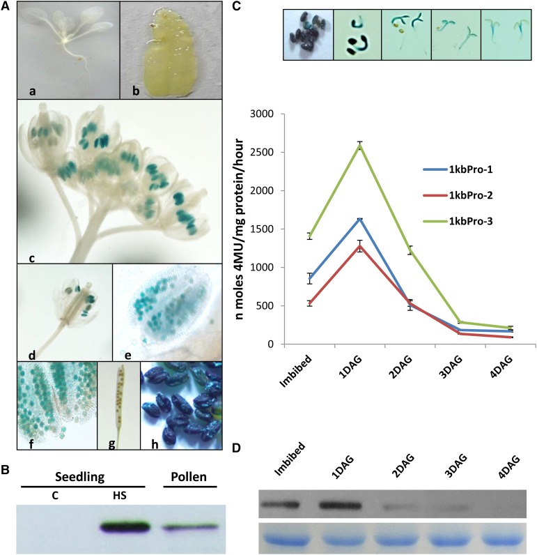
Histochemical staining for Gus expression analysis at different developmental stages of 1kbpro plants under unstressed (control) conditions. A, Ten-day-old seedlings (a), mature leaf from 1-month-old plant (b), inflorescence (c), single flower (d), anther (e), pollen grains (f), young silique (g), and mature imbibed seeds (h). B, Western blot showing the levels of AtClpB-C proteins in pollen of Arabidopsis. Control and heat-stressed (38°C/2 h) protein samples from seedlings were loaded as controls. Two micrograms of total protein from seedlings and 10 µg of total protein from pollen were loaded. C, Gus activity at different stages of seed germination. The graph shows the quantitative estimation through the 4-methylumbelliferyl-beta-d-glucuronide assay, whereas the photograph shows the histochemical Gus staining. D, Western-blot analysis showing the levels of AtClpB-C protein during seed germination. Five micrograms of protein was analyzed, and the bottom row represents the Coomassie Blue-stained protein bands showing equal loading. C, Control; DAG, days after germination; HS, heat stressed. [See online article for color version of this figure.]

