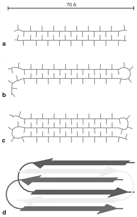
Figure 10.5.
Sketches of the proposed polyglutamine fibril model derived from solid-state NMR data. An approximate scale bar is indicated at the top. Minimal repeat units of (a) polyQ15, (b) polyQ38, and (c) polyQ54 fibrils, assuming similar β-strand lengths, viewed down the fibril axis. (d) Illustration of the superpleated antiparallel cross-β arrangement of monomers in polyQ38 fibrils, viewed down the fibril axis. The fibril repeat unit consists of one GK2Q38K2 molecule that forms two β-strands and contributes to two stacked β-sheets. (Reproduced with permission from Schneider et al. 2011)

