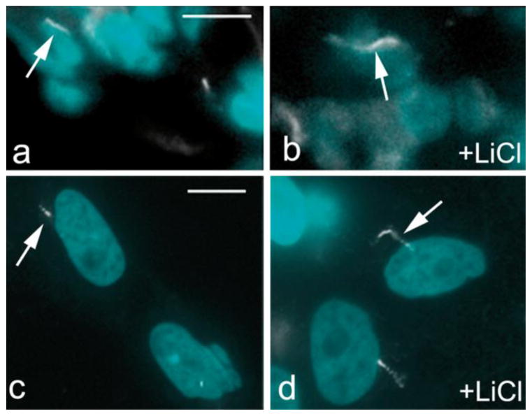
Fig. 3.
Pig testis primary cilia elongate in response to lithium treatment. (a and b): control and lithium treated (+LiCl) pig testis tissue; (c and d): control and lithium treated (+LiCl) Sertoli cell in vitro. Cilia are stained with anti-acetylated tubulin antibody (white; arrows); nuclei are stained with DAPI. The bar indicates 50 μm

