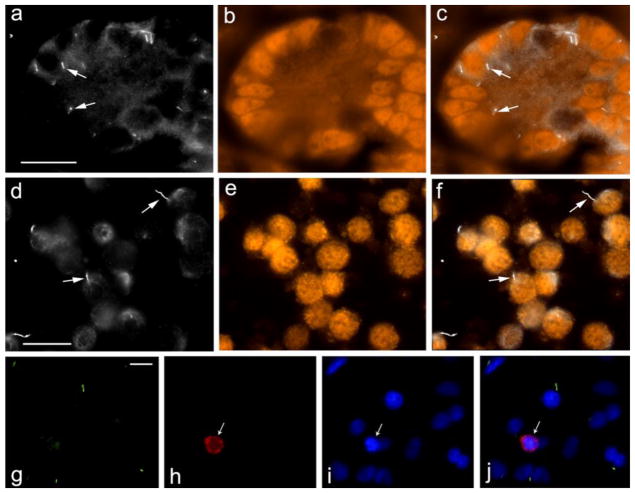
Fig. 4.
Pig Sertoli cells express primary cilia. Pig testis sections (a – c) and cultured Sertoli cells (d – f) were stained with anti-acetylated tubulin antibody to detect primary cilia (a, d; arrows point to examples) and anti-GATA4 antibody to identify Sertoli cells (b, e). c and f show a merged images of a, b and d, e, respectively. The bar indicates 20 μm
Six day pig testis cells, cultured in vitro, were stained with anti-Arl13b antibody to detect primary cilia (g), anti-VASA to detect germ cells (h), DAPI (i). j shows a merged image of g, h, i. The bar indicates 10 μm

