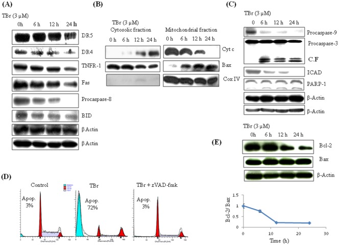
Figure 2. TBr induced apoptosis in HL-60 cells involves both intrinsic and extrinsic apoptotic pathways.

(A) TBr caused the activation of caspase-8 and its downstream target Bid independent of death receptors. HL-60 cells were treated with TBr at 3 µM concentration for indicated time periods. Cells were collected and protein lysates were prepared for imunoblotting of indicative proteins as described in material and methods section. β actin was used as a loading control. (B) TBr treatment triggered the cytosolic translocation of Cyt c from the mitochondria, and mitochondrial translocation of Bax from cytosol. CoxIV was used as a control for mitochondrial fraction purity. (C) TBr caused the activation of caspase-9 followed by caspase-3 and PARP-1 cleavage. β actin was used as an internal control. (D) Cell cycle analysis of TBr (3 µM) treated cells for 24 h along with pan caspase inhibitor z-VAD-fmk (30 µM). Caspase inhibitor was added 1 h before TBr treatment. Cells were collected and stained with PI to determine DNA fluorescence by flow cytometery. (E) Bcl-2/Bax ratio in TBr treated cells in a time dependent manner.
