
Figure 5. TBr induced p-ERK dependent apoptosis in HL-60 cells.
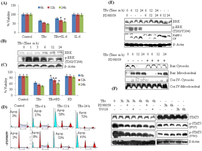
(A) IL-6 treatment increased cell viability in TBr treated cells. Cells were pre-treated with IL-6 (50 ng/ml) for 40 min followed by TBr (3 µM) addition for another 24 h. MTT dye was added 3 h before the experiment termination. Data are mean ± SD from three experiments. (B) TBr (3 µM) induced p-ERK expression in HL-60 cells (C) MTT assay of TBr treated cells along with ERK inhibitor PD98059 (30 µM) at indicated time periods. PD98059 was added 1 h before TBr addition. (D) Effect of PD98059 on subG1 population in TBr treated cells. After treatment, cells were stained with PI and DNA fluorescence was determined by flow cytometery. (E) Effect of PD98059 on Bax and PARP-1 cleavage in TBr treated HL-60 cells. Cells were treated with TBr (3 µM) and PD98059 (30 µM) at indicated time intervals. Whole, mitochondrial and cytosolic protein lystaes were prepared and equal amount of proteins were loaded and resolved on SDS PAGE followed by immunoblotting as described in material and methods section. (F) Effect of PD98059 (10 µM) and U0126 (10 µM) on STAT3 expression in TBr treated cells. β actin was used as a positive control. Data are mean ± SD of three similar experiments and statistical comparisons of TBr vs. TBr with PD98059/IL-6, was made by using the Bonferroni method, *p<0.05, **p<0.01.