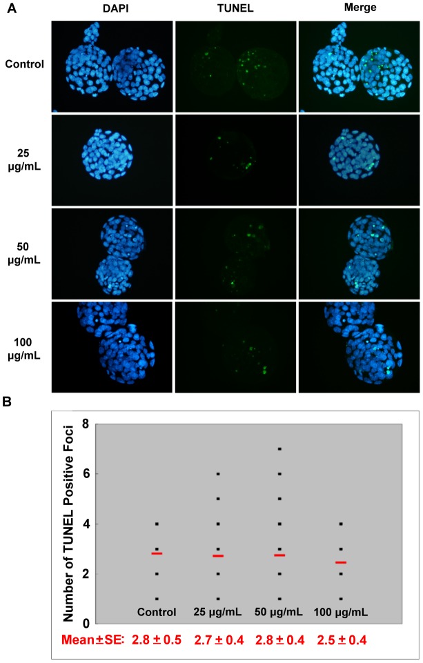
Figure 4. The apoptotic cells in mouse blastocysts.
Blastocysts were derived from pronuclear embryos cultured in different doses of NSP. (A) The nuclei in the blastocyst were stained by DAPI, and the incidence of apoptosis was detected by TUNEL assay. (B) The average number of TUNEL-positive cells in each treatment group is indicated by the short horizontal bar.

