
FIG. 5.
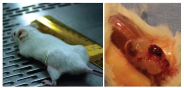
Method for estimating tumor xenograft dimensions in the flank after delivery of U-CH1 cells in a Matrigel carrier. Tumor appears on the mouse flank (left). Large tumor at time of excision exhibits the physaliphorous appearance of the native tumor (right).