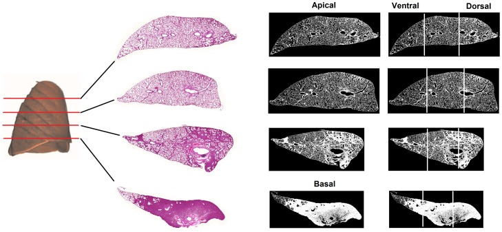
Figure 2. Air fraction obtained by histology images.
Air fraction analysis of apex and base (A) and ventral and dorsal lung areas (B) in the groups: VCV_E (volume-controlled mode and PEEPminErs; n = 6), VCV_E+2 (volume-controlled mode and PEEPminErs plus 2 cmH2O; n = 6), VCV_E-2 (volume-controlled mode and PEEPminErs minus 2 cmH2O; n = 6), VV_E (variable ventilation and PEEPminErs; n = 6), VV_E+2 (variable ventilation and PEEPminErs plus 2 cmH2O; n = 5), VV_E-2 (variable ventilation and PEEPminErs minus 2 cmH2O; n = 6). Ventilatory mode and PEEP effects were tested with two-way ANOVA followed by Tukey post hoc. t test was used to detect differences between ventilated and non-ventilated groups and then correction by comparisons ensued. Statistical significance was accepted at p<0.05. *p<0.05.

