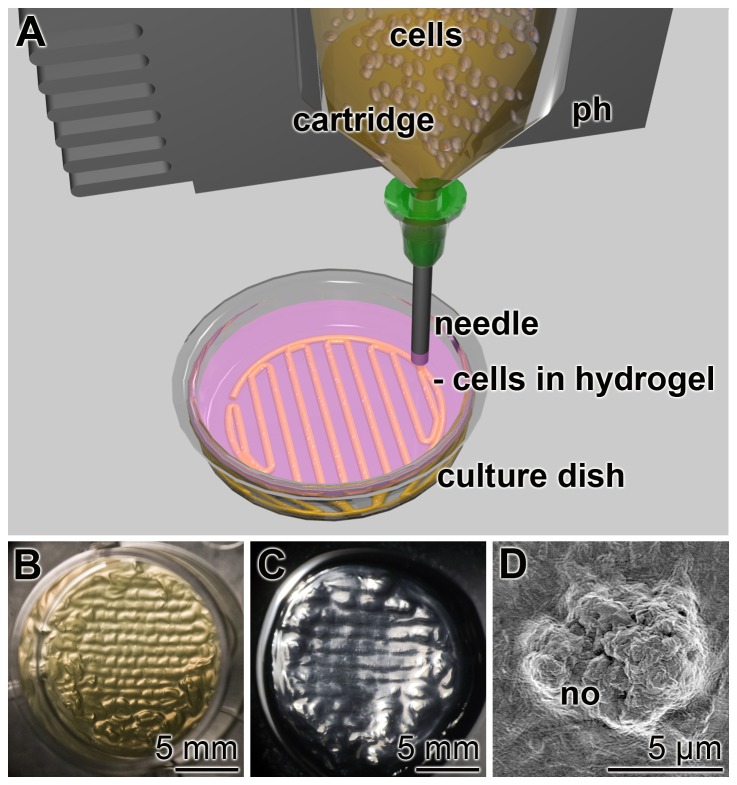
Figure 1. 3D cell bioprinting.
(A) Sketch of the procedure. The alginate/gelatin/SaOS-2 cell suspension is filled into a cartridge, fixed to the printing head (ph). The suspension is pressed through a needle into a culture dish filled with 0.4% CaCl2 as cross-linking solution. (B) A bioprinted stack of 13 mm in diameter and 1.5 mm in height, placed in a 24-well plate. (C) A bioprinted stack after being incubated in medium/FCS for 3 d. (D) SEM image of mineral nodules (no) on the surface of SaOS-2 cells embedded in alginate/gelatin and 100 µmoles/L polyP·Ca2+-complex and incubated for 3 d in the absence and then for 5 d in the presence of the osteogenic cocktail. The specimen was then inspected by SEM.

