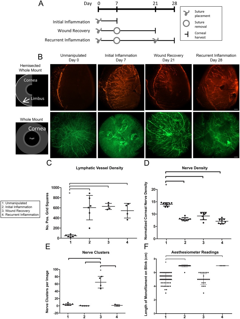
Figure 1. Corneal model of initial inflammation, wound recovery, and recurrent inflammation.
A. Schematic depicts timing of corneal suture placement and removal to induce four distinct tissue microenvironmental conditions: healthy unmanipulated, initial inflammation, wound recovery, and recurrent inflammation. B. Schematics illustrate two corneal mounting styles: hemisected whole mount and whole mount. Images are 100x epifluorescence photomicrographs of corneas immunostained for Lyve-1 (orange) and β-III tubulin (green) and harvested at the indicated time points. Top panel shows Lyve-1+ lymphatic vessels. Bottom panel shows β-III tubulin+ corneal nerves. Scale bars are 100 µm. C. Lymphatic vessel density was quantified from images like those shown in (B). D. Nerve density was quantified from images like those shown in (B). E. Corneal nerve clusters were quantified in wound-recovered tissue. F. A Cochet-Bonnet corneal aesthesiometer was used to measure corneal sensitivity.

