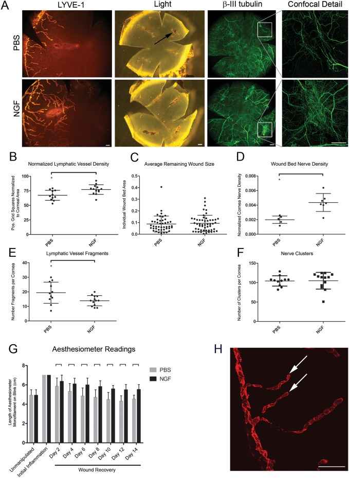
Figure 3. Effects of NGF administration during wound recovery on lymphatic vessel regression, nerve density at corneal wounds, and corneal sensitivity.
After removing sutures to stimulate wound recovery, mouse β-NGF or PBS control was injected subconjunctivally every other day for two weeks. Corneas were harvested, immunostained for Lyve-1 and β-III tubulin, and analyzed by whole mount microscopy. A. Epifluorescence images of Lyve-1+ lymphatic vessels (left panel, scale bar = 100 µm, 100x), bright field images showing remaining wound beds at sites of suture placement (center panel, scale bar = 200 µm, 32x), epifluorescence images of β-III tubulin+ nerves (right panel, scale bar = 200 µm, 32x). Expanded detail shows confocal analysis of β-III tubulin+ nerves at wound beds (offset panel far right, scale bar = 100 µm, 200x). B. Analysis of effects of exogenous NGF administration on corneal lymphatic vessel density during wound recovery from images like those in (A). C. Quantification of average remaining wound size following administration of NGF or PBS. D. Nerve density at remaining wound beds quantified from confocal immunofluorescence images. E. Quantification of lymphatic vessel fragments discontinuous with limbal lymphatic vessel. F. Quantification of nerve clusters in wound-recovered cornea following administration of NGF or PBS. G. Measurements of corneal sensitivity throughout wound recovery period with administration of NGF or PBS. H. 200x confocal immunofluorescence micrograph showing lymphatic vessel fragments (arrowheads). Scale bar = 100 µm.

