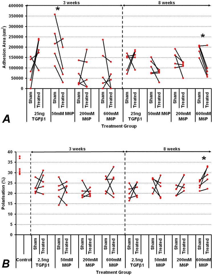
Figure 1. Effect of M6P on tendon adhesion area and tendon structural organisation.
A. Effect of M6P at increasing doses on adhesion area at 3 weeks (most synthetic period) and 8 weeks (end of tendon healing). B. Effect of M6P at increasing doses on tendon organisation as measured by quantitative polarisation. Effect of TGFβ-1 at 2.5 ng was used as a positive control but did not significantly increase adhesion formation or improve tendon organisation (p>0.05). All values of treated tendons were compared to contralateral saline controls. Unwounded controls were also measured for polarisation normalisation. Significant reductions in adhesion formation were seen in 50 mM M6P treated group at three weeks and in 600 mM M6P treated groups at eight weeks. Improvement in tendon organisation was significant with 600 mM M6P treatment. Significant differences with p<0.05 on paired t-testing are indicated by an asterisk.

