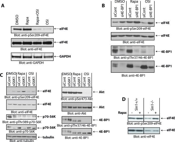
Figure 2. Rapamycin-induced phosphorylation of eIF4E.
(A) Daoy cells were incubated with rapamycin (25 nM) and/or OSI-027 (10 μM) for 90 min. Cell lysates were resolved by SDS-PAGE and immunoblotted with antibodies against the phosphorylated form of eIF4E (pSer-209) or GAPDH (upper and lower panels). Equal amounts of cell lysates from the same experiment were analyzed in parallel by SDS-PAGE and immunoblotted against eIF4E (middle panel). (B) Daoy cells were transfected with control or 4E-BP1 siRNA. After 48 hours, cells were treated with rapamycin (20 nM) or OSI-027 (5 μM) for 90 min. Cell lysates were resolved by SDS-PAGE and immunoblotted with antibodies against the phosphorylated forms of eIF4E (pSer-209) or 4E-BP1 (pThr-37/46). The same blot was stripped and reprobed with antibodies against eIF4E or 4E-BP1, as indicated. (C) Daoy cells were transfected with either control or p70-S6K1 siRNA. After 48 hours, cells were treated with rapamycin (20 nM) or OSI-027 (5 μM) for 90 min. Cell lysates were resolved by SDS-PAGE and immunoblotted with antibodies against the phosphorylated forms of eIF4E (pSer-209), p70-S6K (pThr-389), Akt (pSer-473), 4E-BP1 (pThr-37/46) or tubulin. The corresponding same blots were stripped and reprobed with antibodies against total eIF4E, p70-S6K, Akt or 4E-BP1, as indicated. (D) Sin1+/+ and Sin1-/- MEFs were treated with Rapamycin (20 nM) for 90 min and equal amounts of protein were processed for immunoblotting with antibodies for phosphorylated eIF4E (pSer-209) (upper panel). The immunoblot with antibodies against total eIF4E protein was from lysates from the same experiment analyzed in parallel by SDS-PAGE (lower panel).

