
Figure 2. Immediate binding of ME-9F1 mAb to endothelial cells.
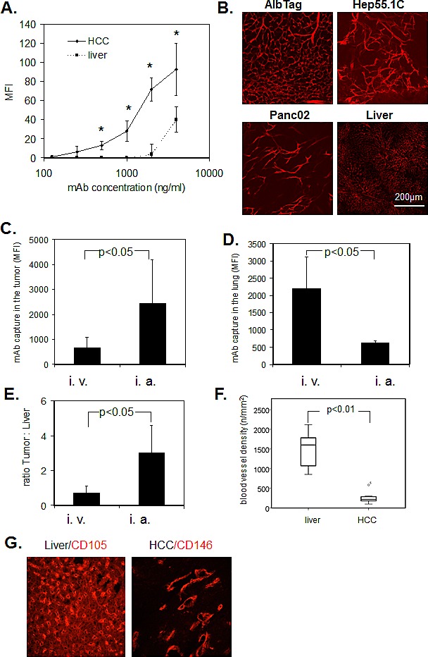
(A) Image-based immunofluorescence analysis of mAb binding to histological slides after 5 s incubation. ME-9F1 showed immediate concentration-dependent binding to tumor endothelial cells, which was significantly higher than binding to hepatic endothelial cells (P<0.05). *Indicates significant differences between tumor and liver tissue. (B) Images of PE-conjugated ME-9F1 mAb binding to tumor endothelial cells in vivo; laser scanning confocal microscopy. Intravenous injection of mAb resulted in excellent visualization of the tumor vascular system in different mouse tumor models. (C–E) Capture of ME-9F1 mAb in tumor and lung tissue after intravenous (i.v.) and intra-arterial (i.a.) injection. mAb binding in tumor (C) and tumor:liver ratio (E) after intra-arterial application was significantly higher than after intravenous injection. Higher mAb binding in the lung was found after intravenous injection (D). (F–G) Blood vessel density (F) and representative images of blood vessel staining in HCC and liver tissue using anti-CD146 or anti-CD105 mAb (G). Tumor blood vessel density in the liver was significantly higher than in HCC from AlbTag mice (P<0.05).