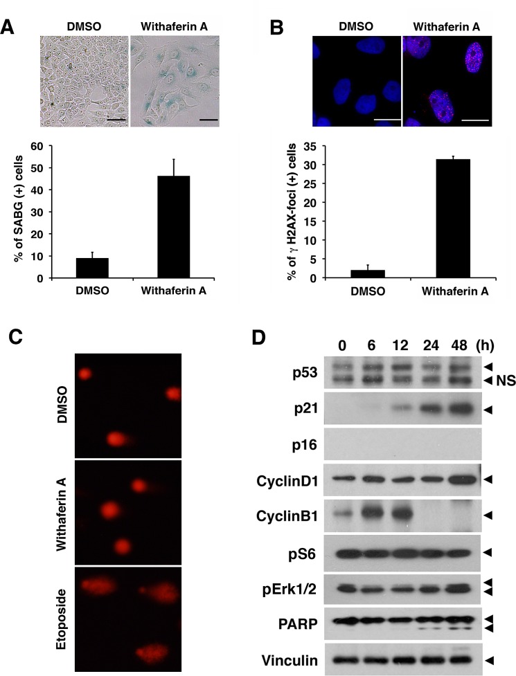
Figure 7. Withaferin A induces cellular senescence and increases p21Cip1 expression in iCSCL-10A cells.
(A) iCSCL-10A cells were treated with DMSO or 1μM of WA for 48 hrs and then stained for senescence-associated b-galactosidase (SABG). Phase contrast microscopy images of the cells are shown (upper). Scale bar, 100 mm. Bars indicate the percentage of SABG-positive cells for each condition and the data shown indicate mean ± SEM (n = 3, lower). One hundred cells per condition were counted and scored. (B) Immunofluorescent analysis of phospho-Histone H2A.X in iCSCL-10A cells treated with DMSO or 1μM of WA for 48 hrs. Nuclei were counterstained with DAPI. Phase contrast microscopy images of the cells are shown (upper). Scale bar, 100 mm. The number of phospho-Histone H2A.X cells was calculated and scored for three independent experiments (lower). Data shown are the mean ± SEM. One hundred cells per condition were counted and scored. (C) Cells were treated as in (B) and subjected to single-cell electrophoresis under denaturing conditions (comet assay). As a positive control, cells treated with10 μg/ml etoposide for 1 hr. (D) Immunoblotting of cell cycle related proteins in iCSCL-10A cells treated with 1 μM of WA at the indicated time points. Vinculin was used as a loading control.

