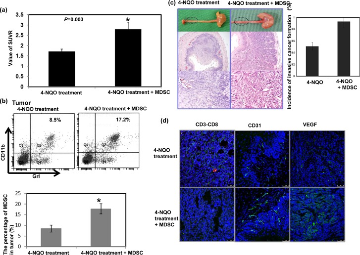
Figure 3. Effect of MDSCs on tumor progression in vivo.
a. The infusion of MDSCs increased the SUVR of PET images in esophageal lesions of mice followed for 12–14 weeks after a 16-week 4-NQO treatment period. The tumor-to-muscle SUVR was calculated from the micro-PET scans. Data points represent the means ± SEMs. *, p<0.05 b. Flow cytometric analysis of CD11b+Gr1+ cells from the 4-NQO-treated mice with or without MDSC infusion. Representative images and quantitative data are shown. The columns represent the means ± SEM. *, p<0.05. c. Representative images of gross lesions and the pathological findings on sectioned tissue samples from the 4-NQO-treated mice with or without MDSC infusion. Quantitative data assessing the incidence of invasive esophageal tumors are shown. The columns represent the means ± SEM. *, p<0.05 d. MDSC infusion attenuated the accumulation of CD3+CD8+ cells (Green: CD3; Red: CD8) and increased CD31 and VEGF immunostaining. Representative images are shown.

