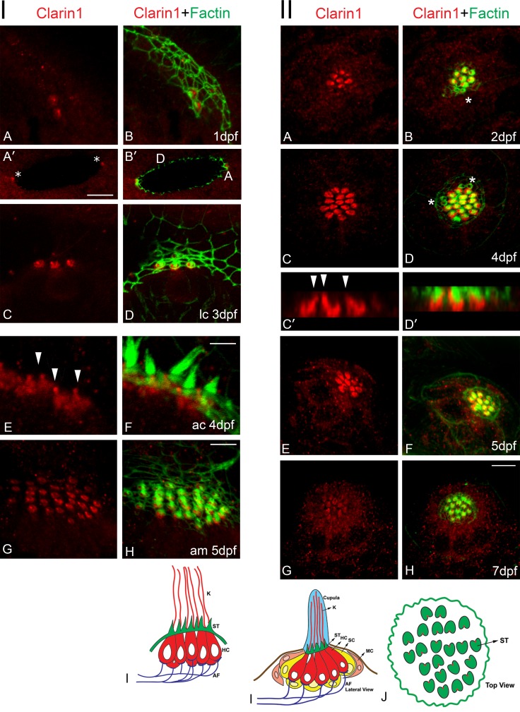
Figure 1.
Clarin-1 protein expression in ear and neuromasts. Confocal images of clarin-1 staining. Actin filaments were counterstained with phalloidin (green). Panel I: (A–B′) 1dpf. A, anterior. D, dorsal. Asterisks in A′ denote macular hair cell precursors. (C–D′) 3 dpf lateral crista (lc). (E and F) 4 dpf anterior crista (ac). Arrowheads in E denote point of insertion of the kinocilium. (G and H) 5 dpf anterior macula (am). (I) Cartoon of a lateral view crista sensory organ. Bars: (A, B, C, D, G and H) 7 µm; (A′ and B′) 16 µm; (E and F) 3 µm. Panel II: (A and B) 2 dpf. (C–D′) 4 dpf. C′ and D′ represent z planes of C and D. Arrowheads in C′ denote point of insertion of the kinocilium. (E and F) 5 dpf. (G and H) 7 dpf. Asterisks denote immature sister hair cells. (I and J) Cartoon of the lateral (I) and top (J) views of a neuromast. K, kinocilia; ST, stereocilia; HC, hair cells; SC, supporting cells; MC, mantle cells; AF, afferent fibers. Results are representative images of >10 independent experiments. Bar, 7 µm.

