Figure 6.
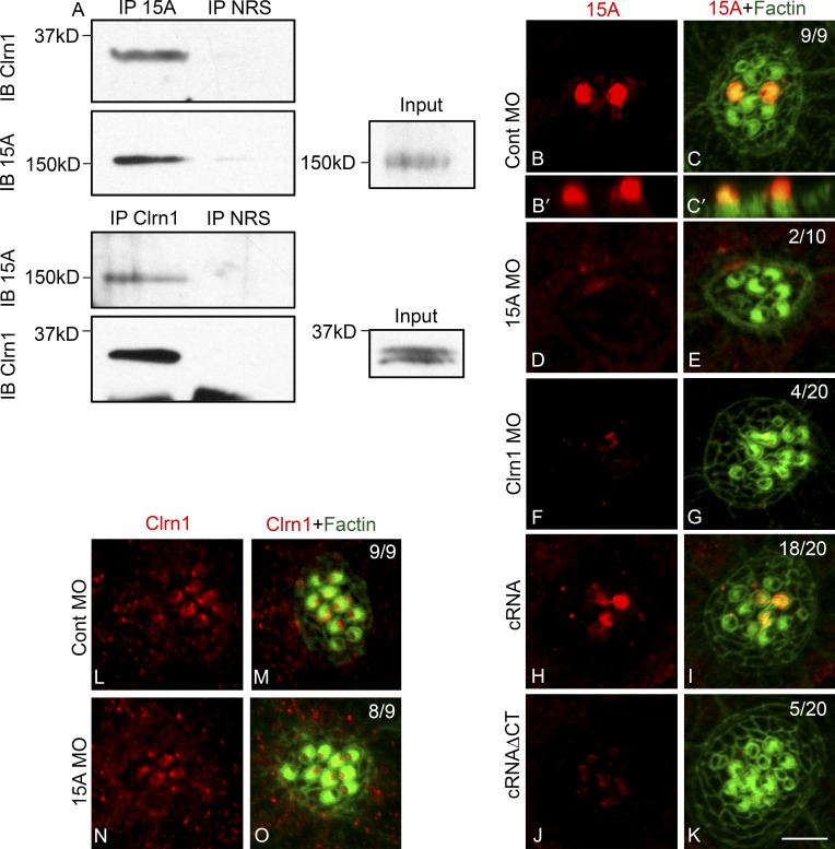
Clarin-1 regulates Pcdh15a distribution. (A) Representative result of three independent experiments of a reciprocal co-IP in 3 dpf larvae. Protein lysates immunoprecipitated with anti-Pcdh15a (IP 15A), anti–clarin-1 (IP Clrn1), or with normal rabbit serum (IP NRS) and immunoblot for clarin-1 (IB Clrn1) or Pch15A (IB 15A). (inputs) 0.5% Pcdh15a and 3% clarin-1. (B–K) Representative images of 3 dpf neuromasts immunostained for Pcdh15a (15A). (B–C′) Pcdh15a control (Cont). (B′ and C′) Z planes of B and C. (D and E) Pcdh15a MOs. (F and G) Clarin-1 MOs. (H and I) MOs + cRNA. (J and K) MOs + cRNAΔCT. (L–O) 3 dpf neuromasts immunostained for clarin-1. (L and M) Pcdh15a control. (N and O) Pcdh15a MOs. Numbers in dual-color images represent number of neuromasts showing normal Usher protein expression/total of neuromasts analyzed in at least nine independent experiments. Bar, 3 µm.

