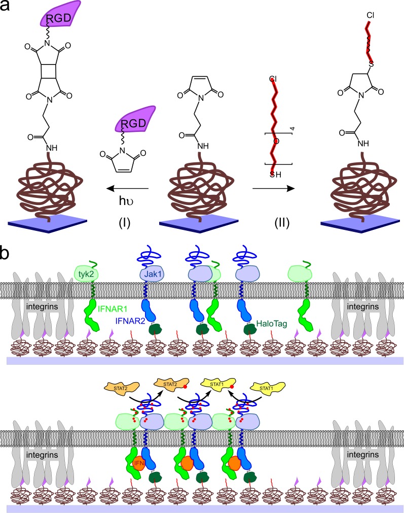
Figure 1.
Strategies for assembly of functional signaling complexes into micropatterns. (a) Binary surface patterning by photochemical coupling of maleimido-RGD to a maleimide-functionalized PEG polymer brush (I) followed by reaction of the nonilluminated maleimide groups with a thiol-functionalized HTL (II). (b) Concept of spatial organization of IFN receptor signaling complexes in the plasma membrane of cells cultured on the surface of a micropatterned coverslide. (top) IFNAR2 fused to the HaloTag is captured into HTL-functionalized areas (HTL functionalities are depicted in red), whereas cell attachment via focal adhesions is mediated by RGD-functionalized areas (RGD functionalities are depicted in violet). (bottom) Upon addition of the IFN, functional complexes are formed by recruitment of IFNAR1 (green), leading to phosphorylation of the associated JAK kinases Jak1 and tyk2 as well as of tyrosine residues on the cytosolic domains of the receptor subunits. Thus, effector proteins such as STAT1 and STAT2 are locally recruited to the receptor.

