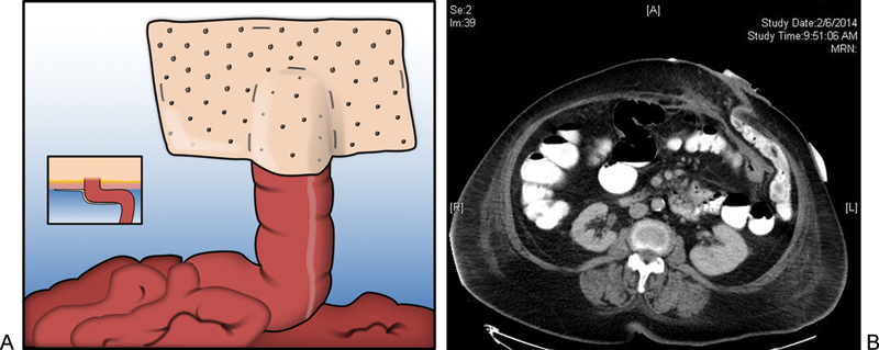
Fig. 1.

(a) Depiction of the Sugarbaker repair. Inset depicts axial view of lateralized bowel traversing abdominal wall with mesh placement relative to bowel and abdominal wall. (b) Postoperative CT scan showing axial view of lateralized bowel traversing the abdominal wall. Note the lateral most portion of bowel as it enters between the biologic mesh and the anterior abdominal wall. Contrast flows freely indicating lack of obstruction.
