
Figure 1. IFN-ε expression in the reproductive tracts of male and female rhesus macaques.
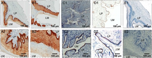
Representative micrographs of IFN-ε expression (stained as brown) in the cervixes, vaginas, and foreskins from at least four SIV-uninfected Indian rhesus macaques detected using IHCS. Five histological sections for each tissue type from each animal were stained. (Upper and lower) The same field at low and high magnifications. IFN-ε expression in the epithelial cells lining the vagina (A1 and A2), ectocervix (B1 and B2), endocervix (C1 and C2), and foreskin (D1 and D2). E1 and E2 show staining of the vaginal tissue with a rabbit IgG isotype as an antibody control. LM, Lumen. Original scale bars: 100 μ.