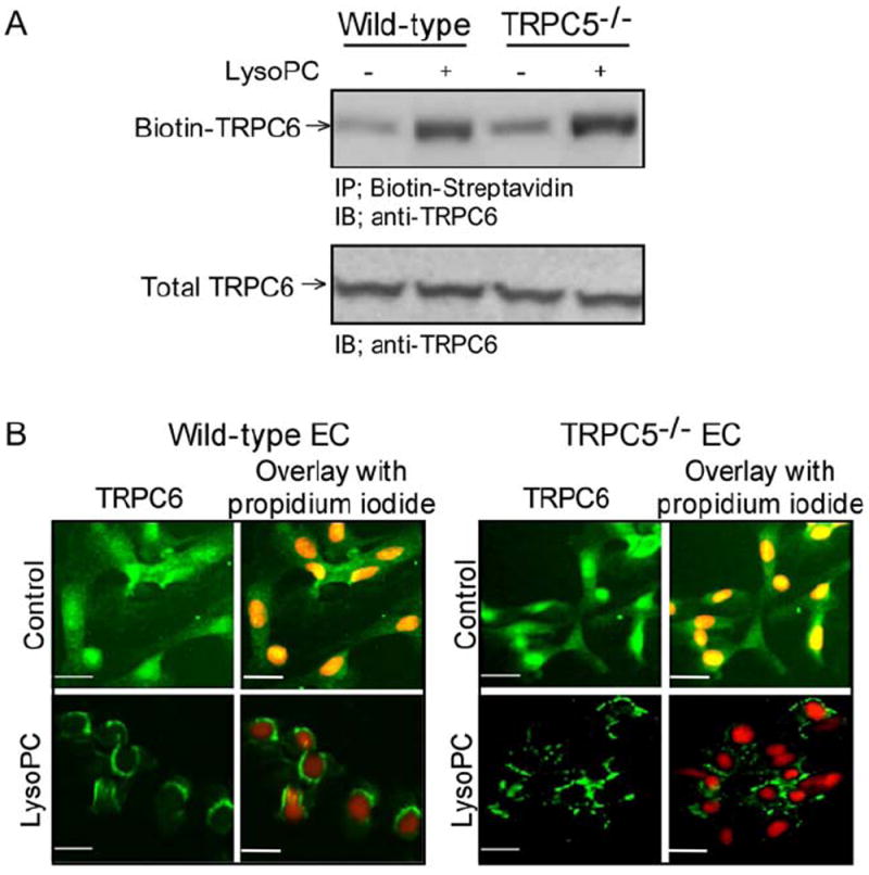
Fig 1. Lysophosphatidylcholine (lysoPC) induces externalization of canonical transient receptor potential (TRPC) 6 protein in TRPC5-/- endothelial cells (ECs).

(A) Wild-type and TRPC5-/- ECs were incubated with or without lysoPC for 1 hour. Cell surface proteins were biotinylated and immunoblot analysis was performed for biotinylated TRPC6 (top panel). Prior to incubation with streptavidin-agarose beads, an aliquot of cell lysate was removed for immunoblot analysis to determine total TRPC6 protein level (bottom panel). (B) WT or TRPC5-/- ECs were incubated with or without lysoPC for 15 minutes, and then exposed to anti-TRPC6 antibody followed by Alexa 488 conjugated secondary antibody. TRPC6 location was assessed by fluorescence microscopy. Nuclei were detected by counterstaining with propidium iodide. Original magnification, x40. Bar, 40 μm.
