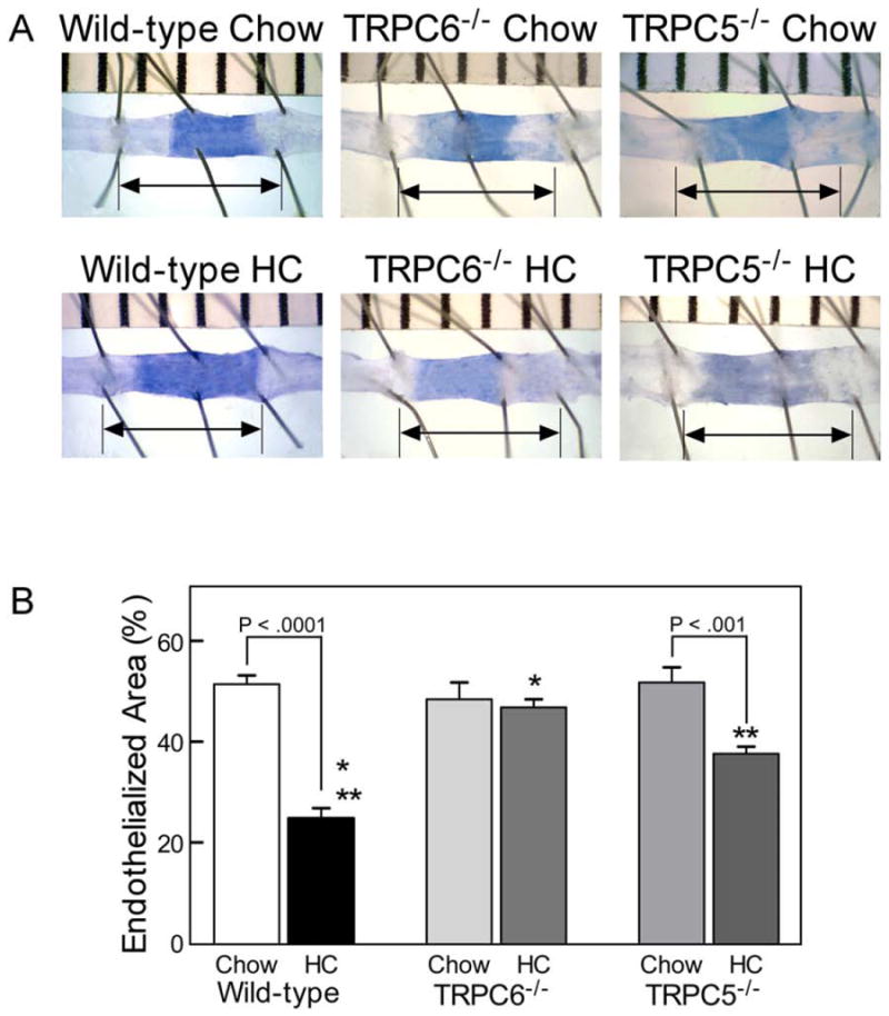
Fig 3. Endothelial healing after carotid artery injury.

(A) Representative images 120 hours after carotid electrocautery. The area without an intact endothelial monolayer stained with Evans Blue. The arrow identifies the length of the original injury. (B) Reendothelialization results shown as the percent of reendothelialized area relative to the total injured area. Results are expressed as the mean ± standard error for each group: wild-type chow diet (n = 10), wild-type high cholesterol (HC) diet (n = 10), TRPC6-/- chow diet (n = 8), TRPC6-/- HC diet (n = 8, * P < .0001 compared with wild-type HC), TRPC5-/- chow diet (n = 8), and TRPC5-/- HC diet (n = 8, ** P = .0001 compared with wild-type HC).
