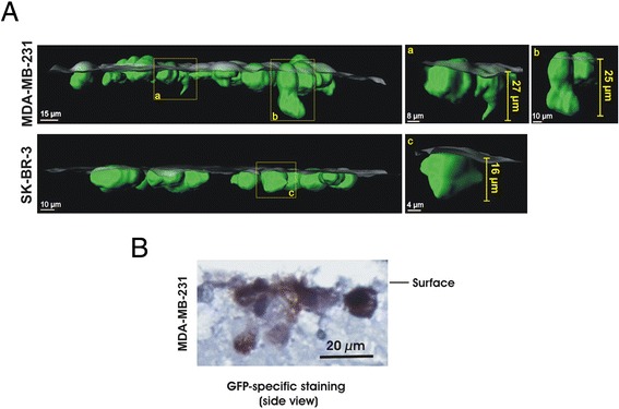
Figure 4.

Application for observation of single cells in PCLS. (A): Partial penetration of MDA-MB-231 or SK-BR-3 cells (green) into PCLS (half transparent grey surface) was analyzed in a side view of IMARIS-generated 3-D images. Yellow frames indicate cancer cells that are extracted from the image to be shown separately (a,b,c). (B): Paraffin sections of PCLS with MDA-MB-231 were stained by GFP specific antibody. Three independent experiments were performed and an example of representative data is shown here.
