Figure 1.

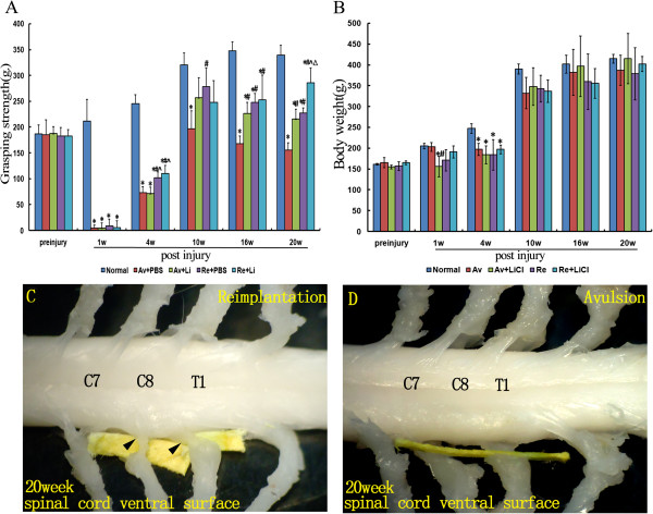
Performance of rats in the grasping test and the gross anatomy of the cervical spinal cord after C7 and C8 ventral root avulsion. Figure 1A shows the average grasping strength of the ipsilateral forepaws in each subgroup of rats at each timepoint before and after the operation and therapies (A). Figure 1B shows the average body weights in each subgroup at each timepoint before and after the operation and therapies. *p < 0.05compared to the normal control group, #p < 0.05 compared to the Av + PBS subgroup, ^p < 0.05 compared to the Av + Li subgroup, and Δp < 0.05 compared to the Re + PBS subgroup (B). Figure 1C and D show the photographs of the gross anatomy of the cervical spinal cord of reimplantation (C) and avulsion (D) animals at the end of the 20 week postinjury period. (C) For reimplantation rats, a nerve was found that was attached close to the ventrolateral surface of the ipsilateral C7 spinal segment and regrown in the middle the trunk of the ipsilateral brachial plexus (arrows). Additionally, the other reimplanted ventral root that originated from the ventral surface of the ipsilateral C8 spinal segmentwas identified regrowing into the lower trunk of the ipsilateral brachial plexus (arrows). (D) An obvious gap between distal part of the C7 and C8 spinal nerves and the ventral surface of the correspondingspinal cord was detected in avulsion animals (yellow stick).
