Figure 4.

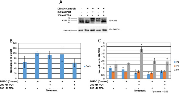
PQ1 changes isoform expression of Cx43. Cells were treated with: no treatment (control), DMSO (control), 200 nM PQ1, 200 nM TPA and 200 nM PQ1 + 200 nM TPA for 1 hour. Level of Cx43 and its isoforms were examined by western blot analysis. GAPDH was used as a loading control. A) Levels of Cx43 were detected using anti-connexin43 (F-7) antibody specific for amino acids 357–381 at the C-terminus domain. B) Graphical presentation of three independent experiments showing pixel intensities of total Cx43 normalized to control. C) Graphical presentation shows the ratio of Cx43 isoforms P0, P1 and P2. Data were obtained in three independent experiments and are represented as the mean ± SD. *P value is <0.05 compared to control. IB = Immunoblot against Cx43.
