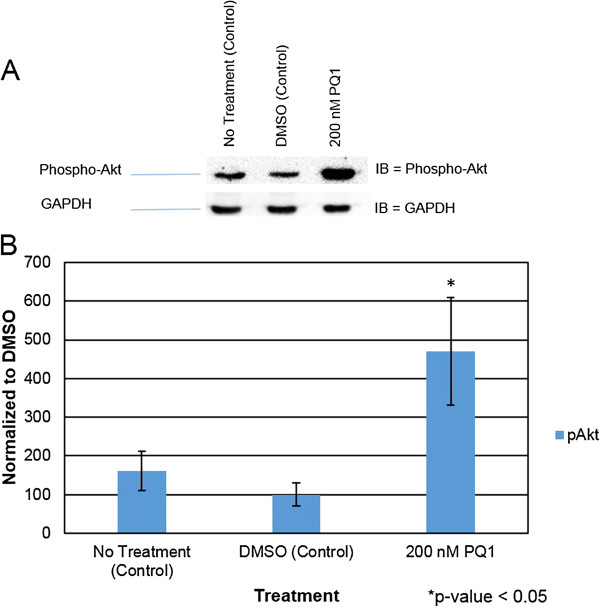
Figure 6.

PQ1 causes activation of Akt. Cells were treated with: no treatment (control), DMSO (control) and 200 nM PQ1 for 1 hour. Level of phospho-Akt (active Akt) was examined by western blot analysis. GAPDH was used as a loading control. A) Level of active Akt was detected using anti-phospho-Akt (Ser473) (D9E) antibody specific for activated Akt. B) Graphical presentation of three independent experiments showing pixel intensities of active Akt normalized to control. *P value is <0.05 compared to control. IB = Immunoblot against phospho-Akt.
