
Figure 3.
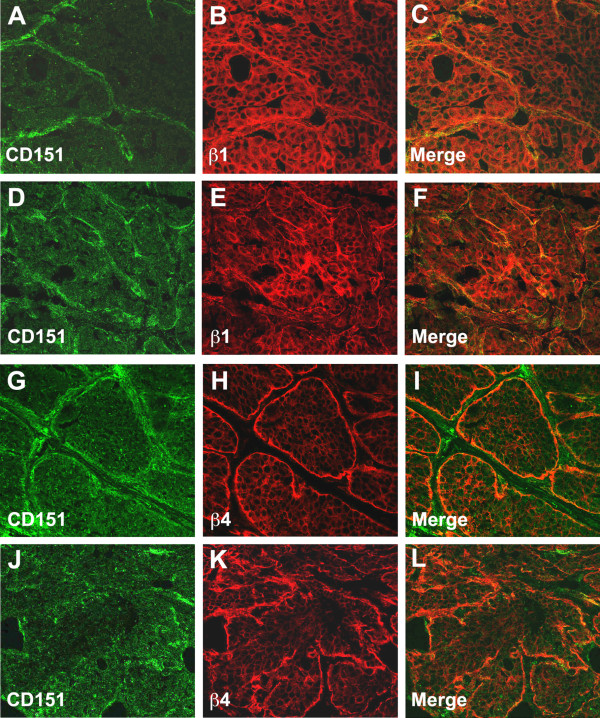
Expression of CD151 in mammary tumors. Dual immunofluorescent labeling and confocal analysis using CD151 antibodies (green) and antibodies towards β1-(A-F), or β4-integrin (G-L) were performed on Cd151+/+ MMTV/PyMT mammary tumors at the adenoma stage (A-C and G-I) and the carcinoma stage (D-F and J-L) (stages as defined by Lin et al. [17]). At the early adenoma stage (A-C and G-I), the tumor cells showed faint CD151 expression overall that was more intense on the edge of the tumor nodules, colocalizing with β1 and β4 integrins along the basement membrane. At a more advanced tumor stage, the labeling was more patchy with still some degree of colocalization with integrins (D-F and J-L). Original magnification ×400.