
Figure 1.
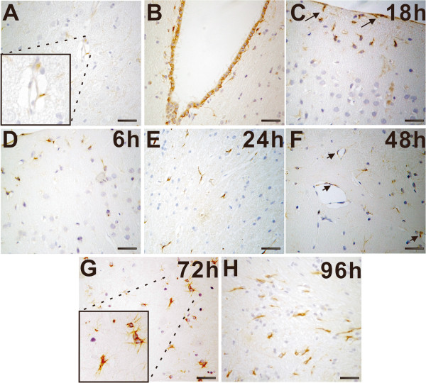
Accumulation of connective tissue growth factor+ (CTGF+) non-neuron cells in traumatic brain injury. A: In normal brains, CTGF immunoreactivity was occasionally observed in the brain tissue. The boxed area indicates CTGF immunoreactivity in small vessels. B: Almost all ependymal cells showed CTGF+. C-H: The accumulation of CTGF+ cells in the lesioned regions at 18 h (C), 6 h (D), 24 h (E), 48 h (F), 72 h (G) and 96 h (H). Arrows indicate the accumulation of CTGF+ non-neuron cells with astrocytes phenotype in the cortex and meningeal (C). CTGF immunoreactivity was also detected in endothelial cells (F). A significant increase in CTGF+ non-neuron cells was observed confined to the lesion at 72 h postinjury (G). The boxed area indicates the localization of CTGF+ non-neuron cells that were mainly identified as astrocytes with typical stellate morphologic characteristics or belt-shaped, grouped pattern. The accumulation of CTGF+ non-neuron cells reached the maximum at 96-h postinjury (H). Scale bars are 50 μm.