
Figure 2.
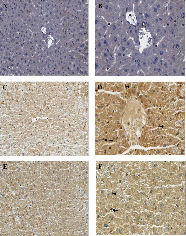
Immunohistochemical staining of IL-12 antibody in the representative tissue specimens. A-B: Control. C-D: APAP. E-F: Aloe vera-treated. Images were obtained at × 20 (A, C, and E) and × 40 (B, D, and F). DAB staining was used to highlight liver Kupffer cells in each section (dark brown stain, arrows).