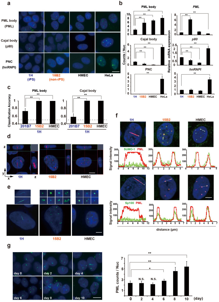
Figure 2. Quantitative assessment of nuclear structures in completely and incompletely reprogrammed human iPSCs.
(a) Identification of nuclear structure characteristics of iPSCs. Immunostaining was performed to identify the PML body (PML), Cajal body (p80 coilin), and perinucleolar compartment (PNC) (hnRNP I) (green). Nuclei were stained with DAPI (blue). (b) Quantification of nuclear structure formation (n>200, left) and the mRNA levels of the corresponding components in the structures (n = 3, right). (c) wndchrm classifications against iPSCs (1H) using immunofluorescence images of PML and Cajal bodies (n = 10). (d) Detection of linear PML structures by three-dimensional confocal microscopy. (e) Detection of PML structural variation by structured illumination microscopy (100 nm resolution). Enlarged images of PML structures are shown in the upper boxes. (f) Lack of SUMO-1 and Sp100 in the linear PML structures of bona fide iPSCs. The signal intensity along the arrow is shown below. PML, red; SUMO-1 and Sp100, green. (g) Transition of PML structures from linear to round during differentiation. The number of PML structures is shown at the right (n>300). Values are the means and s.d. *, p<0.05; **, p<0.01. Scale bars, 5 μm.

