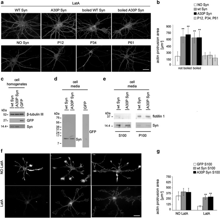
Figure 2.
Unfolded and released wt and A30P Syn affect actin cytoskeleton similarly to the purified protein. (a) F-actin distribution of 14 DIV embryonic hippocampal neurons incubated without or with 1 μM purified wt or A30P Syn, either untreated or boiled; or with 1 μM Syn synthetic peptides (P12, P34, P61). Neurons were treated with LatA and stained as in Figure 1a. (b) Quantitative evaluation of actin protrusion areas of neurons treated as in a, calculated as in Figure 1b. (c) Immunoblots of cell homogenates from 2 DIV hippocampal neurons electroporated with coding constructs for either green fluorescent protein (GFP) or wt or A30P Syn. β-Tubulin III is shown as an internal standard. (d, e) Immunoblots showing the presence of Syn in the culture media collected from 2 DIV hippocampal neurons electroporated as in c (d), and in their soluble and vesicular fractions (e) separated by ultracentrifugation (S100 and P100, respectively). Flotillin 1 is shown as a marker of exosomal membranes. (f) F-actin distribution in 1 DIV hippocampal neurons incubated ON with S100 fractions from GFP-, wt Syn- or A30P Syn-expressing neurons; neurons were treated and stained as in Figure 1a. (g) Quantitative evaluation of actin protrusion areas of neurons treated as in f, calculated as in Figure 1b. In b and g, data are expressed as mean values ±S.D.; in b, n=30 neurons from two independent cultures, in g, n=60 neurons from three independent cultures. Statistical significance was determined by one-way ANOVA followed by Dunnett's test for multiple comparison, **P<0.01. Bar in a and f: 20 μm

