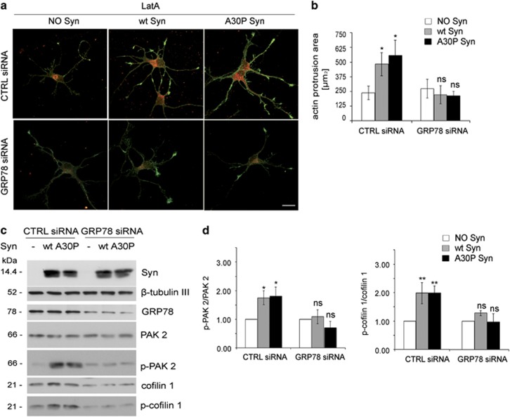
Figure 6.
GRP78 downregulation prevents the effects triggered by extracellular wt and A30P Syn. (a) Fluorescence analysis of embryonic hippocampal neurons electroporated at 1 DIV with GRP78 siRNA sequences or with not-targeting siRNA sequences (CTRL siRNA), incubated at 4 DIV for 1 h in the absence or presence of 1 μM purified wt or A30P Syn and then treated for 1 h with 1 μM LatA. Neurons were processed for fluorescent phalloidin staining (F-actin, green) and indirect immunofluorescence of GRP78 (red). (b) Quantitative evaluation of actin protrusion areas in neurons treated as in a. Actin protrusion areas were calculated by subtracting β-tubulin III projection areas from F-actin projection areas. (c) Representative immunoblots of cell homogenates from hippocampal neurons electroporated at 1 DIV with GRP78 siRNA sequences and incubated at 4 DIV in the absence or presence of purified wt or A30P Syn as in a. β-Tubulin III is shown as an internal standard. (d) Quantification of the ratio between p-PAK 2 and total PAK 2 (left panel) and p-cofilin 1 and total cofilin 1 (right panel) bands, analyzed by densitometry, for each experimental condition in c; values are normalized to those of control samples not incubated with Syn (NO Syn). In b and d, data are expressed as mean values ±S.D. In b, n=60 neurons from three independent cultures; in d, n=3 independent experiments. Statistical significance was determined by one-way ANOVA followed by Dunnett's test for multiple comparison, *P<0.05; **P<0.01; ns=not significant. Bar in a: 20 μm

