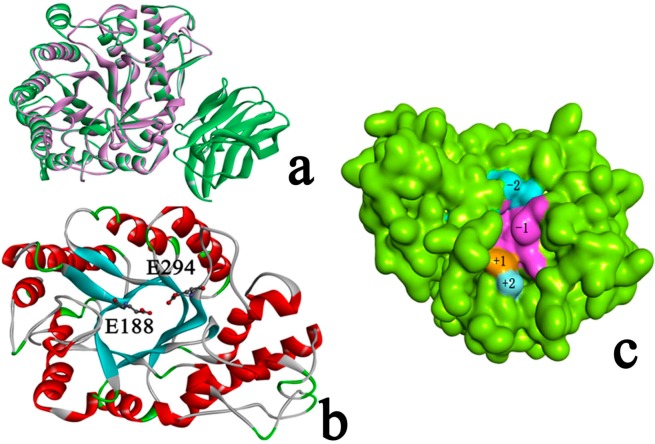
Figure 4.
(a) Superimposition of Xyn10A (purple) and the template Xyn10b (green) (RMSD 0.32 Å); (b) The 3D structure of Xyn10A. Glu188 and Glu294 located at the end of β-strands 4 and 7, repetitively; (c) The surface of Xyn10A. Color light blue represents for subsite +2, color brown represents for subsite +1, color purple represents for subsite −1, and color dark blue represents for subsite −2.

