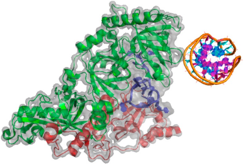
Figure 3.
Structure of the DNA G-quadruplex of an Oxytricha nova telomeric protein-DNA complex (PDBid: 1JB7) [50]. Sugar-phosphate backbone and nucleobases in the DNA quadruplex structure is depicted by the orange ribbon and purple/cyan cartoons, respectively. The α- and β-subunits of the G-quadruplex-binding protein are represented by the green and red cartoons, respectively. A single-stranded DNA is represented by the blue cartoon. Grey color highlights the electron cloud of the protein-DNA complex.

