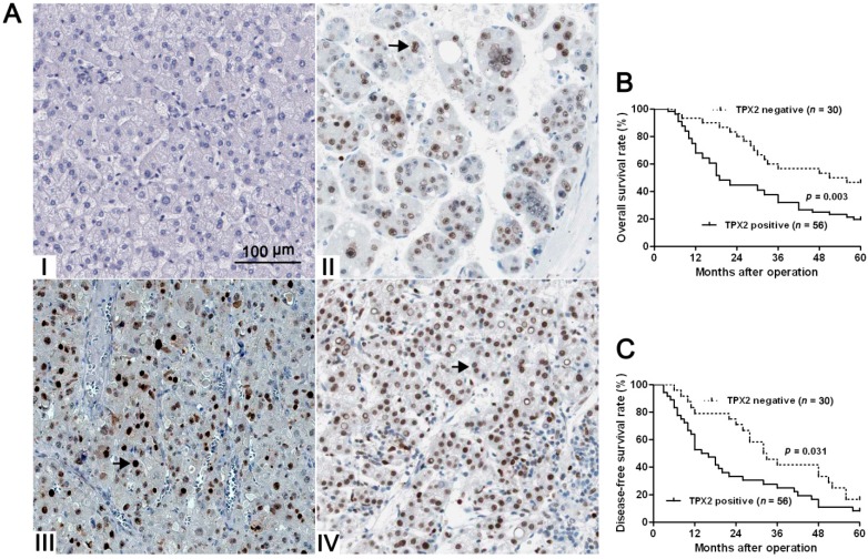
Figure 2.
The immunostaining of TPX2 and its prognostic significance in HCC specimens. (A) Immunohistochemical staining of TPX2 in HCC. TPX2 was localized within the nuclei. Elevated expression of TPX2 (the arrows) in the tumor cells of HCC tissue (II, III, and IV) compared to normal tumor-adjacent tissues with negative staining (I) (scale bar: 100 μm); (B) The overall survival (OS) and (C) disease-free survival rates (DFS) were estimated by the Kaplan-Meier method. Both the OS rate and DFS rate of patients with TPX2 positive primary tumor were significantly lower than that of patients with TPX2 negative primary tumor (log-rank test, p < 0.05).

