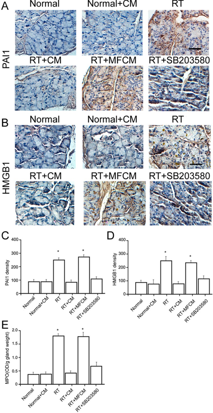
Figure 3.
iPSC-CM suppressed the RILI-associated inflammatory response. (A) Immunohistochemical staining for PAI1 in iPSC-CM treated RILI and normal mouse lacrimal glands. Scale bar = 25 µm; (B) Immunohistochemical staining for HMGB1 in iPSC-CM treated RILI and normal mouse lacrimal glands. Scale bar = 25 µm; (C,D) Quantification of the mean density of immunohistochemical staining of these sections (CM: iPSC-CM, RT: radiotherapy, MFCM: mouse fibroblasts conditioned medium). N = 5. Values are means ± SEM. * p < 0.01; (E) Neutrophils migrated into the injured gland sites revealed by the neutrophil counts and myeloperoxidase (MPO) assay. * p < 0.01.

