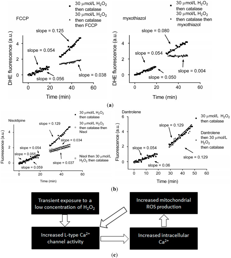
Figure 1.
Transient exposure to H2O2 leads to L-type Ca2+ channel-dependent increase in mitochondrial superoxide, triggering ROS-induced-ROS release. (a) dihydroethidium (DHE) fluorescence recorded from individual guinea pig cardiomyocytes before and after exposure to 30 µM H2O2 for 5 min followed by 10 U/mL catalase for 5 min then 2 nM FCCP (an uncoupler of oxidative phosphorylation) as indicated (left panel) or 7 nM myxothiazol (to block electron transport at mitochondrial complex III) (right panel); (b) DHE fluorescence recorded from individual guinea pig cardiomyocytes before and after exposure to 30 µM H2O2 for 5 min followed by 10 U/mL catalase for 5 min and the L-type Ca2+ channel antagonist nisoldipine (nisol; 2 µM) as indicated (left panel) or ryanodine receptor antagonist dantrolene (20 µM; right panel); and (c) Schematic illustrating the persistent increase in intracellular ROS and intracellular Ca2+ following a transient H2O2 exposure or ROS-induced ROS-release. Reproduced with permission from [40].

