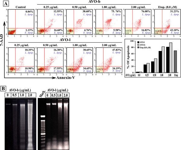
Figure 2.

The growth inhibitory effect of AVO on HL-60 cells is mediated by apoptosis. (A) Cultured HL-60 cells were treated with varying concentration of AVO-b or AVO-l (0.0 to 2.0 μg/mL) an apoptosis was analyzed by a flow cytometer after staining with FITC-annexin-V and 7-AAD. The scattered blots showing the percentages of early and late apoptosis are indicated for one experiment. The graph represents the summary mean percentages of apoptosis (early and late apoptosis) of two independent experiments for each concentration of the oil. HL-60 cells were treated with 8.0 μM etoposide as a positive control. Statistical analysis showed that all samples are significantly different (p < 0.05), when compared to untreated controls. (B) Genomic DNA was extracted from HL-60 cells treated with the different concentrations of AVO-l or AVO-b, and applied to agarose gel electrophoresis. DNA fragmentation in the gel was visualized by UV light after staining with ethidium bromide. Mark, is a DNA ladder marker.
