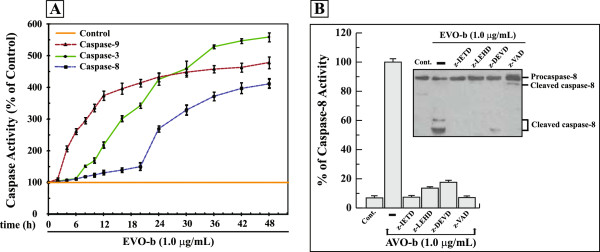
Figure 4.

AVO-b-induced processing and activation of caspase-8 in HL-60 cells requires activation of caspase-9 and -3. (A) HL-60 cells were treated with 1.0 μg/mL AVO-b for different time intervals (0 to 48 h), and caspase-9 (▲), caspase-8 (■) and caspase-3 (●) activities were determined calorimetrically and expressed as relative percentages to the control untreated cells. The data shown represent the means ± SD of two independent trials for each time point. (B) AVO-b-induced caspase-8 activation in the presence and absence (-) of 35 μM specific caspase-8 inhibitor (z-IETD-fmk), caspase-9 inhibitor (z-LEHD-fmk), caspase-3 inhibitor (z-DEVD-fmk), or 50 μM of the general caspase inhibitor (z-VAD-fmk). HL-60 cells were incubated with each of the individual caspase inhibitor for 6 hours before the exposure to 1.0 μg/mL AVO-b for an additional 24 h. Caspase-8 activation was analyzed calorimetrically after addition of the substrate Ac-IETD-pNA (the graph) and by immunoblotting with a specific anti-caspase-8 antibody (the Western blot). The results shown represent the percentages of caspase-8 activity relative to the control cells treated with AVO-b alone (-). “Cont.”, represents caspase-8 activity in HL-60 of untreated cells. Results of caspase-8 activity represent the means ± SD of three indecent trials. Statistical analysis showed that all samples treated with specific caspase inhibitors and AVO-b are significantly different (p < 0.05), when compared to cells treated with AVO-b alone.
