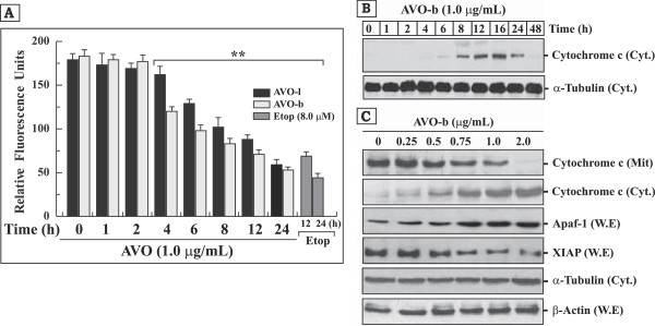
Figure 5.

AVO induces disruption of mitochondrial transmembrane potential (ΔΨm), promotes the release cytochrome c to the cytoplasm and modulates the expression of Apaf-1 and XIAP proteins in HL-60 cells. (A) HL-60 cells treated with 1.0 μg/ml AVO-l or AVO-b for different periods of time (0–24 h) and changes in mitochondrial membrane potential was monitored by a flourimetric analysis after addition of the fluorescent stain JC-1. Cells treated with 8.0 μM etoposide for 12 and 24 h were used as positive controls. The results shown represents the mean ± SD of three independent experiments. **Represents samples that are statistically different, when compared to their controls at 0 time or AVO untreated cells. (B) Western blot analysis of cytochrome c in the cytosolic fraction (50 μg) of HL-60 cells treated with AVO-b for different time points (0 to 48 h) as indicated at the top of each lane. (C) Western blot analysis of Apaf-1 and XIPA in whole cell extracts (W.E; 50 μg), and cytochrome c in cytosolic (Cyt.; 50 μg) and mitochondrial (Mit.; 30 μg) fractions obtained from HL-60 cells treated with varying concentrations of AVO-b (0.0–2.0 μg/mL). The same Western blots in both (B) and (C) for the cytosolic fractions and whole cell extracts were probed with antibodies to α-tubulin and β-actin, respectively, to ensure equal protein loading in the different lanes.
