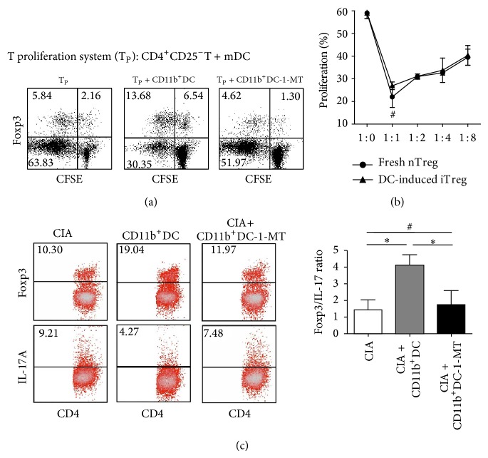
Figure 5.
The conversion of Foxp3+Tregs from CD4+CD25−T cells through coculture with isolated splenic CD11b+IDO+DCs. (a) CD4+CD25−T cells (105) stimulated with mDCs (2 × 10⁴) were cocultured for 4 days with isolated splenic CD11b+DCs (104) with or without 1-MT pretreatment. The cells were surface-stained with anti-CD4 mAbs, followed by intracellular staining with anti-Foxp3 mAbs to determine the frequency of CD4+Foxp3+Tregs. The dates are presented using a dot plot, expressed as the % positive cells. The results are representative of three experiments showing similar results. (b) After cocultivation for 4 days, as described in (a), CD11b+DC-induced CD4+CD25+T cells were isolated and used as inhibitors at different doses to suppress the proliferation of new CD4+CD25−T cells in response to stimulation with anti-CD3/28 mAbs. The suppressive activity of freshly isolated CD4+CD25+T cells was also examined as a control. The proliferative responses were measured after 4 days. The data are representative of three experiments with similar results, reported as the means ± SEM. # P > 0.05 compared with the indicated groups using unpaired t-tests. (c) CD4+T cells from the spleens of CIA mice treated with CD11b+DC with or without 1-MT on the third week after the onset of arthritis were collected and intracellularly stained with anti-Foxp3 and anti-IL-17A mAbs. FACS was used to measure the percent frequency of positively stained cells, and the frequencies of Treg/Th17 cells are expressed as the means ± SEM of four independent experiments. # P > 0.05, * P < 0.05 compared with the indicated groups using unpaired t-tests.

