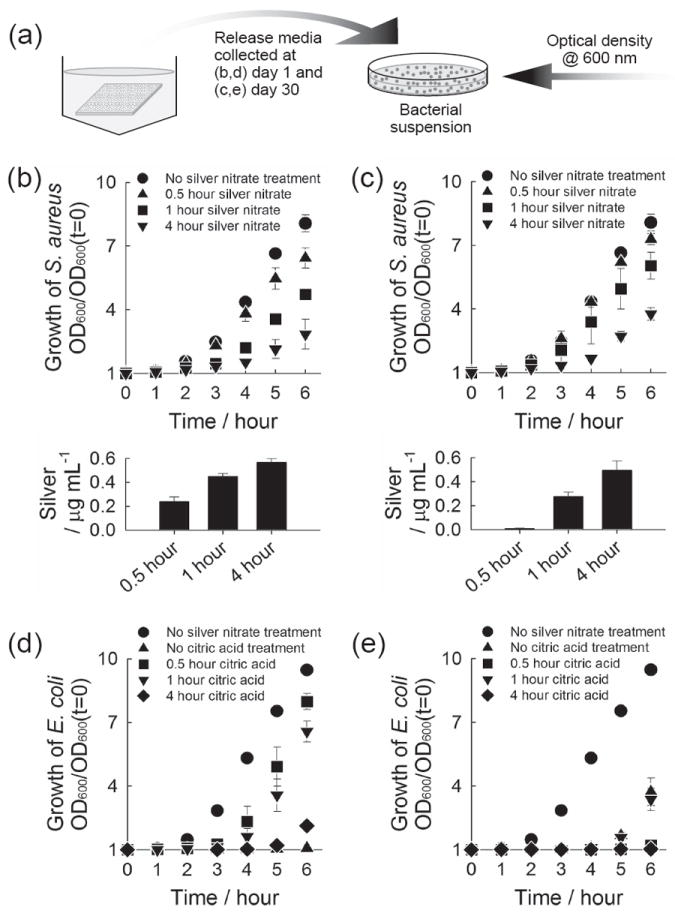
Figure 5.

Antibacterial activity of released silver. (a) Schematic of experiment. (b-e) Growth curve of S. aureus (b,c) and E. coli (d,e) upon the addition of release media. The release media were collected from CaP coatings which were incubated sequentially in (b,c) 5 mm citric acid solution for 1 hour and 5 mm silver nitrate solution for different time periods, and (d,e) 5 mm citric acid solution for different time periods and 5 mm silver nitrate solution for 1 hour. The release media collected (b,d) at day 1 where CaP coatings were incubated for the first day and (c,e) at day 30 where the coatings were incubated from day 20 to day 30, were added to S. aureus (b,c) and E. coli (d,e). Lower plots in (b) and (c) show the concentration of silver in the bacteria solution tested.
