
Figure 4.
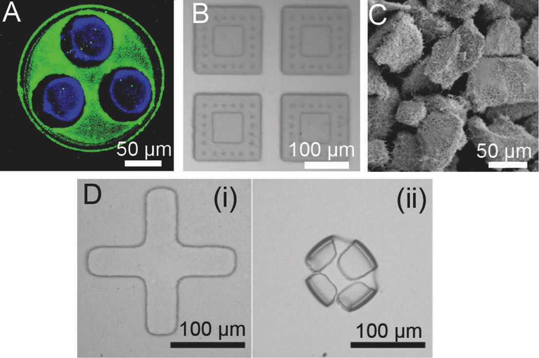
Chemical and structural modifications to enhance microdevice adhesion and increase residence time. A. Fluorescence micrograph of FITC-lectin (green) asymmetrically coated onto the drug-releasing side of microdevices for targeted bioadhesion to the intestinal mucosa with fluorescently labeled BSA shown in blue [34]. B. Microdevices with microposts designed to penetrate the mucus membrane surrounding a drug reservoir [57]. C. Planar nanowire-coated microparticles dramatically increase surface area and enhance microdevice adhesion through increased non-covalent interactions [76]. D. Self-folding microdevices shown before (i) and after (ii) exposure to water are designed to mechanically attach to intestinal tissue [60]. Reproduced with permission.