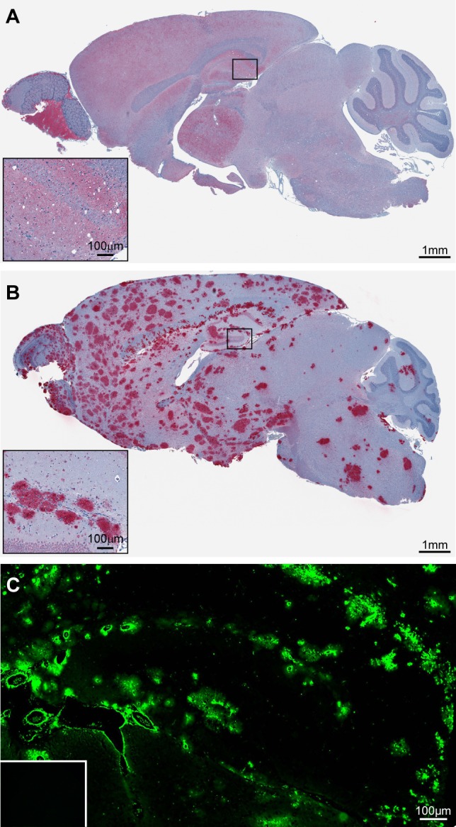
Figure 1.

Infection of C57BL/10 and Tg44 mice with the RML prion strain yields two distinct disease phenotypes. (A) Immunostaining of RML infected C57BL/10 brain tissue with D13 prion antibody shows diffuse PrPSc deposition in most regions of the brain in addition to spongiform degeneration. (B) Immunostaining of RML infected Tg44 brain tissue with D13 prion antibody shows PrPSc deposition in the absence of spongiform degeneration. (C) Staining of the brain hippocampal region with the amyloid-specific fluorescent dye Thioflavin S confirms the presence of amyloid plaque in RML infected Tg44 brain. Thioflavin S staining is negative in uninfected Tg44 mice (inset).
